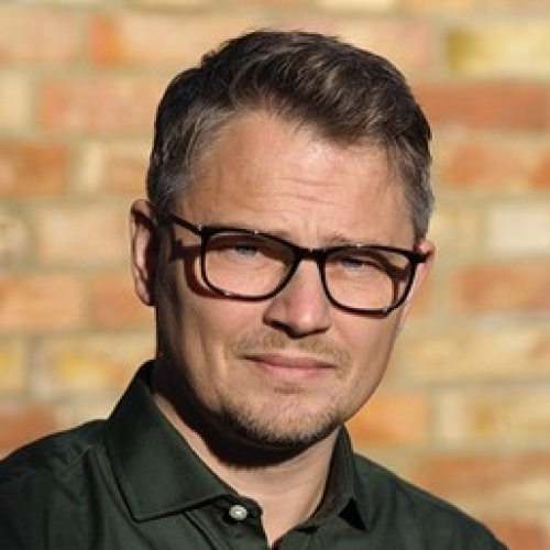
Dr Lothar Schermelleh
Born and raised in Munich, Germany, I studied Biosciences at the local Ludwig Maximilian University (LMU). In 2003, I obtained my doctorate in Molecular Cytogenetics under the mentorship of Thomas Cremer, a pioneer of in situ hybridisation and fluorescence microscopy techniques and renowned for his discovery of chromosome territories. As a visual person interested in design and architecture, and being captivated by the secrets of life, his scientific vision of nuclear architecture as a critical contributor to mammalian genome function struck a chord and motivated me to pursue a career in academia.
My early work on the cell cycle-dependent dynamic organisation of chromosomes required pushing the boundaries of live cell 3D microscopy and developing new and improved fluorescence labelling methods. This was only possible in close partnership with physicists and optical engineers, imprinting in me the value of interdisciplinary collaboration as a route to break new ground.
Following this theme, I later joined the Molecular Epigenetics group of Heinrich Leonhardt as a Postdoctoral Researcher and Lecturer, where I focused on studying the role and regulation of DNA methyltransferase 1 (Dnmt1) through developing novel single-cell imaging and quantitative analysis approaches. During this time, as a visiting scientist in the lab of John W. Sedat at the University of California San Francisco, I first encountered super-resolution structured illumination microscopy (SIM) and later established this exciting imaging technique for a broad range of cell biological applications.
In 2011, I joined the University of Oxford as Micron Senior Research Fellow and Principal Investigator at the Department of Biochemistry, where my group advanced the development of quantitative super-resolution 3D imaging to study mesoscale chromatin organisation and mechanisms of epigenetic gene regulation, e.g. in X chromosome inactivation. My long-term aspiration is to literally “see our genes and genomes at work”, in molecular detail using ever more elaborate super-resolution fluorescence and correlative 3D cryo-electron microscopic approaches.
In 2020, I was named Associate Professor and became Academic Director of the Micron Bioimaging Facility. After spending more than ten years of research at Oxford University, I am incredibly excited and grateful to join Pembroke College as a Senior Associate and contribute to (and learn from) a thriving and diverse community of outstanding scholars and students.
01/2020 – present Associate Professor; Department of Biochemistry, University of Oxford;
08/2011 – 12/2019 Micron Senior Research Fellow, Principal Investigator; Department of Biochemistry, University of Oxford
08/2003 – 07/2011 Postdoctoral Researcher / Lecturer (Epigenetics & Bioimaging); Faculty of Biology, Ludwig Maximilian University Munich (Advisor: Prof Heinrich Leonhardt)
2003 Doctor of Science, with highest honour (Dr. rer. nat. summa cum laude) Ludwig Maximilian University Munich (Advisor: Prof Thomas Cremer)
1998 Diploma in Biology, Ludwig Maximilian University Munich
Selected recent publications:
Brown JM, De Ornellas S, Parisi E, Schermelleh L, Buckle VJ. 2022. RASER-FISH, a non-denaturing fluorescence in situ hybridization for preservation of three-dimensional interphase chromatin structure. Nat Protoc 17, 1306-1331.
Rodermund L, Coker H, Oldenkamp R, Wei G, Bowness J, Rajkumar B, Nesterova T, Pinto DMS, Schermelleh L#, Brockdorff N#. 2021. Time-resolved structured illumination microscopy reveals key principles of Xist RNA spreading. Science 372(6547), eabe7500.
Miron E, Oldenkamp R, Brown JM, Pinto DMS, Xu CS, Faria AR, Shaban HA, Rhodes JDP, Innocent C, de Ornellas S, Hess H, Buckle V, Schermelleh L. 2020. Chromatin arranges in chains of mesoscale domains with nanoscale functional topography independent of cohesin. Science Advances 6, eaba8811.
Ochs F, Karemore G, Miron E, Brown JM, Sedlackova H, Rask M, Lampe M, Buckle V, Schermelleh L#, Lukas J#, Lukas C. 2019. Stabilization of chromatin topology safeguards genome integrity. Nature 594(7779): 571-574.
Schermelleh L#, Ferrand A, Huser T, Eggeling C, Sauer M, Biehlmaier O, Drummen GPC#. 2019. Super-resolution microscopy demystified. Nat Cell Biol 21(1): 72-84.
Demmerle J, Innocent C, North AJ, Ball G, Müller M, Miron E, Matsuda A, Dobbie IM, Markaki Y, Schermelleh L. 2017. Strategic and practical guidelines for successful structured illumination microscopy. Nat Protoc. 12(5): 988-1010.
Dr Lothar Schermelleh

Born and raised in Munich, Germany, I studied Biosciences at the local Ludwig Maximilian University (LMU). In 2003, I obtained my doctorate in Molecular Cytogenetics under the mentorship of Thomas Cremer, a pioneer of in situ hybridisation and fluorescence microscopy techniques and renowned for his discovery of chromosome territories. As a visual person interested in design and architecture, and being captivated by the secrets of life, his scientific vision of nuclear architecture as a critical contributor to mammalian genome function struck a chord and motivated me to pursue a career in academia.
My early work on the cell cycle-dependent dynamic organisation of chromosomes required pushing the boundaries of live cell 3D microscopy and developing new and improved fluorescence labelling methods. This was only possible in close partnership with physicists and optical engineers, imprinting in me the value of interdisciplinary collaboration as a route to break new ground.
Following this theme, I later joined the Molecular Epigenetics group of Heinrich Leonhardt as a Postdoctoral Researcher and Lecturer, where I focused on studying the role and regulation of DNA methyltransferase 1 (Dnmt1) through developing novel single-cell imaging and quantitative analysis approaches. During this time, as a visiting scientist in the lab of John W. Sedat at the University of California San Francisco, I first encountered super-resolution structured illumination microscopy (SIM) and later established this exciting imaging technique for a broad range of cell biological applications.
In 2011, I joined the University of Oxford as Micron Senior Research Fellow and Principal Investigator at the Department of Biochemistry, where my group advanced the development of quantitative super-resolution 3D imaging to study mesoscale chromatin organisation and mechanisms of epigenetic gene regulation, e.g. in X chromosome inactivation. My long-term aspiration is to literally “see our genes and genomes at work”, in molecular detail using ever more elaborate super-resolution fluorescence and correlative 3D cryo-electron microscopic approaches.
In 2020, I was named Associate Professor and became Academic Director of the Micron Bioimaging Facility. After spending more than ten years of research at Oxford University, I am incredibly excited and grateful to join Pembroke College as a Senior Associate and contribute to (and learn from) a thriving and diverse community of outstanding scholars and students.
01/2020 – present Associate Professor; Department of Biochemistry, University of Oxford;
08/2011 – 12/2019 Micron Senior Research Fellow, Principal Investigator; Department of Biochemistry, University of Oxford
08/2003 – 07/2011 Postdoctoral Researcher / Lecturer (Epigenetics & Bioimaging); Faculty of Biology, Ludwig Maximilian University Munich (Advisor: Prof Heinrich Leonhardt)
2003 Doctor of Science, with highest honour (Dr. rer. nat. summa cum laude) Ludwig Maximilian University Munich (Advisor: Prof Thomas Cremer)
1998 Diploma in Biology, Ludwig Maximilian University Munich
Selected recent publications:
Brown JM, De Ornellas S, Parisi E, Schermelleh L, Buckle VJ. 2022. RASER-FISH, a non-denaturing fluorescence in situ hybridization for preservation of three-dimensional interphase chromatin structure. Nat Protoc 17, 1306-1331.
Rodermund L, Coker H, Oldenkamp R, Wei G, Bowness J, Rajkumar B, Nesterova T, Pinto DMS, Schermelleh L#, Brockdorff N#. 2021. Time-resolved structured illumination microscopy reveals key principles of Xist RNA spreading. Science 372(6547), eabe7500.
Miron E, Oldenkamp R, Brown JM, Pinto DMS, Xu CS, Faria AR, Shaban HA, Rhodes JDP, Innocent C, de Ornellas S, Hess H, Buckle V, Schermelleh L. 2020. Chromatin arranges in chains of mesoscale domains with nanoscale functional topography independent of cohesin. Science Advances 6, eaba8811.
Ochs F, Karemore G, Miron E, Brown JM, Sedlackova H, Rask M, Lampe M, Buckle V, Schermelleh L#, Lukas J#, Lukas C. 2019. Stabilization of chromatin topology safeguards genome integrity. Nature 594(7779): 571-574.
Schermelleh L#, Ferrand A, Huser T, Eggeling C, Sauer M, Biehlmaier O, Drummen GPC#. 2019. Super-resolution microscopy demystified. Nat Cell Biol 21(1): 72-84.
Demmerle J, Innocent C, North AJ, Ball G, Müller M, Miron E, Matsuda A, Dobbie IM, Markaki Y, Schermelleh L. 2017. Strategic and practical guidelines for successful structured illumination microscopy. Nat Protoc. 12(5): 988-1010.